Kristina M Kemp, MD FRCP(C)
University of Manitoba
Winnipeg, Manitoba
AND
Kenneth Roberts, MD FRCP(C)
Southlake Regional Health Centre
Newmarket, Ontario
AND
Susan M Tarlo, MB BS FRCP(C)
Gage Occupational and Environmental Health Unit,
Professor of Medicine
University of Toronto
Toronto, Ontario
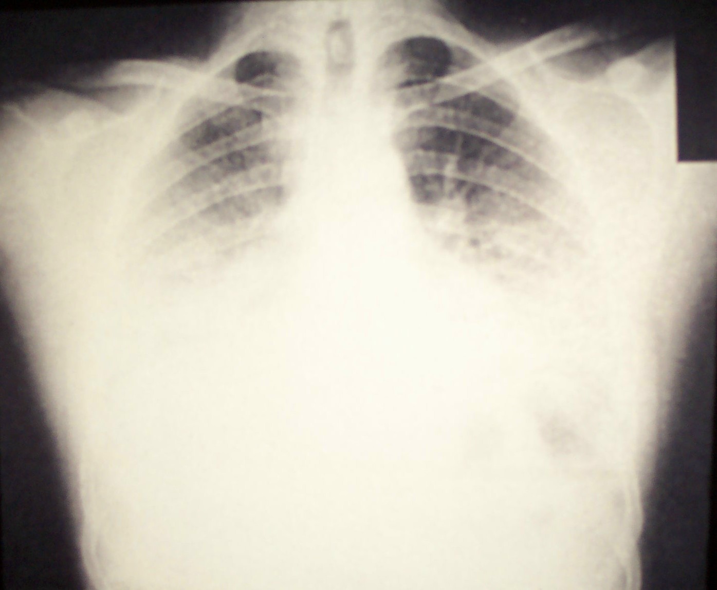
A 24-year-old, life-long nonsmoking man worked for five years as a foreman in a plant where carrots and onions were cleaned and packed. He was seen in the emergency department with a 1-week history of dry cough, dyspnea, and mild fever. Clinical examination revealed a temperature of 38.2? C, tachypnea, tachycardia (120/min) and crackles, mainly basal, on auscultation of his chest. He was found to have severe hypoxemia (PO2 50 mm Hg, O2 saturation 85% on room air) and elevated white blood cell count (16.1 x 10⁹/L) with neutrophilia and a left shift. Chest radiography revealed diffuse bilateral infiltrates (Fig. 1). He was admitted to the intensive care unit, and rapidly improved with supplemental oxygen, intravenous fluids and antibiotics. On day 5, his chest radiograph was clear and he was discharged home.
Click on images to enlarge.
Figure 1: Chest radiograph at initial presentation demonstrating diffuse bilateral infiltrates
The patient experienced similar symptoms 2 days after return to work, and was readmitted to hospital with similar findings. He improved within 12 hours, clearing within 3 days with supportive management only, without antibiotics or corticosteroids.
Both episodes occurred after working 8 hours in the onion room, soon after installation of plastic curtains to maintain heat in the area. He returned to work in the separate carrot room and had no symptoms, except after revisiting the onion room on two occasions, when symptoms recurred 3 hours following even short visits.
Spirometry at the time of symptoms showed restrictive changes, clearing between episodes (Table 1). Serum precipitins were identified from the patient against Aspergillus flavus and Aureo pullulans. Fungal culture from uncleaned onions from the workplace showed heavy growth of A. Aspergillus fumigatus, various penicillium species, and a lighter growth of A. flavus and A. fusarium species.
A diagnosis of onion worker’s hypersensitivity pneumonitis (HP) was made, due to fungal exposure at work. The patient moved to a different workplace and had no further symptoms.
Table 1: Pulmonary function test results| FEV1 (% predicted) | FVC (% predicted) | DLCO (% predicted) | |
| During episode | 2.7 L (63) | 2.8 L (54) | Not done |
| Post episode | 4.5 L (104) | 5.2 L (102) | 94 |
Figure 2: Carrots and onions

COMMENTS: Please send any comments to Akshay Sood, M.D., M.P.H.
SUGGESTED RESOURCES:
REFERENCES:
- Tarlo S. Is your patient’s workplace causing lung disease? Can J Diagnosis 2001; 18:74-85.
- Rose CS. Hypersensitivity pneumonitis. In: Murray JF, Nadel JA, editors Textbook of respiratory medicine, 3rd ed..Philadelphia: W. B. Saunders Company; 2000. pp.1867 – 1884.
- Richerson HB, Bernstein IL, Fink JN, et al. Guidelines for the clinical evaluation of hypersensitivity pneumonitis Report of the subcommittee on hypersensitivity pneumonitis. J Allergy Clin Immunol 1989; 84(5): 839-844.
- Patel AM, Ryu JH, Reed CE. Hypersensitivity pneumonitis: Current concepts and future questions. J Allergy Clin Immunol 2001; 108(5): 661-670.
- Schuyler M, Cormier Y. The Diagnosis of hypersensitivity pneumonitis. Chest 1997; 111(3): 534-536.
- Lacasse Y, Selman M, Costabel U, et al. Clinical diagnosis of hypersensitivity pneumonitis. Am J Respir Crit Care Med 2003; 168(8): 952-958.
- Stevens DA, Kan VL, Judson MA, et al. Infectious disease Society of America Practice guidelines for diseases caused by Aspergillus. Clinl Infect Dis 2000; 30:696-709.
- Cormier Y, Desmeules M. Treatment of hypersensitivity pneumonitis (HP): comparison between contact avoidance and corticosteroids. Can Respir J 1994; 1:223-8.
Question 1
This patient's differential diagnosis from initial history alone does not include which ONE of the following?
- Acute pneumonia
- Acute endotoxin exposure effects
- Organic dust toxic syndrome
- Reactive airways dysfunction syndrome
- Occupational asthma
- Idiopathic pulmonary fibrosis
Answer
The correct answer is F.
As this case illustrates, a good occupational history improves diagnostic considerations. This case was initially misinterpreted as acute pneumonia due to similarity in the presenting symptoms (1).
Other important clinical syndromes can occur as a result of inhalation of organic agents. Acute exposure to endotoxin in bio-aerosols is strongly associated with respiratory symptoms, airflow limitation, and longitudinal declines in airflow among agriculture and industrial workers. Organic dust toxic syndrome (ODTS) is a nonimmunologic reaction associated with microbial contamination of organic dust, which occurs more commonly than hypersensitivity pneumonitis (HP) in farmers. ODTS is associated with intense exposure occurring on a single day, whereas HP is associated with high exposure on most days for prolonged periods.
Occupational asthma and reactive airway dysfunction syndrome (RADS) should be considered in this patient given the exposure at the workplace. RADS occurs after a single exposure to an irritant and persists beyond 3 months. Occupational asthma and RADS can be differentiated from HP by spirometric evaluation, as an obstructive pattern is shown in the former two and a restrictive pattern in the latter.
Idiopathic pulmonary fibrosis (IPF) would not be considered in this patient’s diagnosis given the acute presentation and rapid resolution. IPF requires symptoms to be presenting for greater than 3 months. In the chronic form, hypersensitivity pneumonitis (HP) may resemble IPF as well as sarcoidosis, bronchiolitis, and BOOP.
Question 2
Which of the following is/are helpful in making a diagnosis of hypersensitivity pneumonitis? (more than one answer may be correct)
- Clinical symptoms of dyspnea and cough
- Interstitial changes on chest radiography
- Serum precipitating antibodies to an offending antigen
- Restrictive pattern with impaired gas exchange on pulmonary function testing
- Bronchoalveolar lavage fluid lymphocytosis with predominant CD8 T-suppressor cells
- Open lung biopsy with diffuse interstitial disease and non-caseating granulomas
Answer
The correct answer is All of the above.
Hypersensitivity pneumonitis is a disease with differing forms of clinical presentation, disease severity, and natural history (2). There is no single test to confirm the diagnosis; therefore, many different diagnostic criteria for HP have been proposed (3-6).
The guidelines for the clinical evaluation of hypersensitivity pneumonitis (3), suggested in a 1989 report, establishes the diagnosis of HP if: (a) the history and physical findings and pulmonary function tests indicate an interstitial lung disease, (b) the x-ray film is consistent, (c) there is exposure to a recognized cause, and (d) there is antibody to that antigen. In other exceptional circumstances, bronchoalveolar lavage may help, but biopsy is rarely needed.
The diagnostic criteria for hypersensitivity pneumonitis, as adapted from Schuyler and Cormier (5), involve both major and minor criteria, mainly designed to differentiate HP from sarcoidosis and ILD. The major criteria include: (a) history of symptoms compatible with hypersensitivity pneumonitis that appear or worsen within hours after antigen exposure, (b) confirmation of exposure to the offending agent by history, investigation of the environment, serum precipitin test, and/or bronchoalveolar lavage fluid antibody, (c) compatible changes on chest radiography or high-resolution computed tomography of the chest, (d) bronchoalveolar lavage fluid lymphocytosis, if bronchoalveolar lavage performed, (e) compatible histologic changes, if lung biopsy performed, (f) positive “natural challenge” or controlled inhalational challenge. Minor criteria include: (a) basilar crackles, (b) decreased diffusion capacity, (c) arterial hypoxemia, either at rest or with exercise. The diagnosis is confirmed if the patient exhibits four of the major criteria and two of the minor criteria. Additionally, other diseases with similar symptoms need to be ruled out.
Lacasse et al. (6) recently developed a clinical prediction rule for the clinical diagnosis of hypersensitivity pneumonitis. Six significant predictors of HP were identified: (a) exposure to a known offending antigen, (b) positive precipitating antibodies to the offending antigen, (c) recurrent episodes of symptoms, (d) inspiratory crackles on physical examination, (e) symptoms occurring 4 to 8 hours after exposure, (f) and weight loss. Using the prediction rule from these variables, the area under the receiver-operating characteristic curve was 0.93, and a table derived from these variables can determine the probability of having HP, such that invasive procedures such as bronchoalveolar lavage and open lung biopsy may be avoided in many cases.Question 3
Which ONE of the following lung diseases is NOT related to Aspergillus exposure?
- Allergic bronchopulmonary aspergillosis (ABPA)
- Allergic fungal sinusitis
- Asthma
- Aspergilloma
- Lung cancer
Answer
The correct answer is E.
Aspergillus species are saprophytic molds found worldwide and can result in allergic and infectious disease in humans (7). Pulmonary Aspergillus infections include aspergilloma and invasive pulmonary aspergillosis. Aspergillus sinonasal infections may be invasive and can follow an indolent to fulminant course. Aspergillus sinus infections may also present as a fungus ball or mycetoma. Aspergillus species are a cause of allergic fungal sinusitis, which typically occurs in immunocompetent, atopic young adults.
Aspergilloma most often occurs within pulmonary cavities or ectatic bronchi in patients with underlying disease such as old tuberculous or histoplasmosis cavities, bullous emphysema, fibrotic lung disease, or fibrocystic sarcoidosis. It can co-exist with ABPA, or with invasive aspergillus pneumonia in immunocompromised hosts.
APBA is a hypersensitivity disease of the lungs most often caused by A. fumigatus. Asthma is included in the diagnostic criteria for ABPA; however, even in the absence of ABPA, inhalation of Aspergillus spores can trigger IgE-mediated bronchospasm.
Aspergillus colonization or infection has been reported to complicate lung cancer especially in cavitary lesions, but no causation of lung cancer has been established by the infection.
Question 4
Appropriate recommendations for this patient include which ONE of the following?
- Advise him that he will progress to end-stage lung disease if he continues to work in the onion room
- Advise him that he may continue at his current workplace if he takes prednisone as therapy
- Advise him to avoid further work in the onion room
- Advise him to have future work environments checked for mold
- Advise him not to eat onions
Answer
The correct answer is C.
The best advice for this patient is complete avoidance of the causative agent. For some, this course of action is not possible due to severe financial or emotional factors. In such circumstances, the next best advice is to use protective devices such as a respirator, which limit exposure to the inciting antigen. The use of corticosteroids is advocated in the initial management of severe cases of hypersensitivity pneumonitis; however placebo-controlled clinical trials are lacking. Systemic steroids are not advocated for management with ongoing chronic exposure, but have been used in chronic farmers’ lung if avoidance is not financially feasible (8).
The prognosis of acute HP is good when appropriate interventions are undertaken. However, even in patients who practice avoidance, some will progress to end-stage lung disease. The corollary is also true where some patients with continued antigen exposure do not progress in their disease. Although it is reasonable to avoid overtly moldy environments, it is unnecessary to have future work areas checked for nonvisible mold, and this should only be considered if symptoms recur.
This patient does not have to avoid onions, as the causative agent was the Aspergillus mold.


