Can "U" see it?
Scott M Brewster, MD
University of Utah, Department of Internal Medicine
Case
Patient is a 59 year old female with past medical history of pulmonary artery hypertension undergoing diuretic adjustment for lower extremity edema. She had successful reduction in edema but was noted to be more confused and lethargic by family, prompting visit to emergency department.
EKG is shown below. What is the diagnosis?
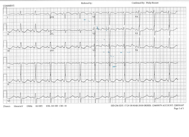
The EKG above shows new onset U waves (positive deflections after the T wave) in V2 and V3. This patient had a recent addition of metolazone and bumetanide to her diuretic regimen. At presentation her potassium was noted to be low at 2.1 mEq/L. Hypokalemia typically presents with three major EKG findings:
- ST-segment depression
- Flattening of T wave and prolonged QT interval
- Presentation of U waves.
U waves are deflections following the T wave, usually best seen in the anterior leads, V2- V3, and typically positive. U waves are seen in hypokalemia, appearing in almost 80% of those with K < 2.7 mEq/L. However, select antiarrhythmic drugs, central nervous system disorders and occasionally healthy hearts will occasionally show U waves as well.
The long QT or QTU in hypokalemia can portend fatal consequences with increased rates of torsades-de-pointes and ventricular tachycardia. The above patient was admitted to the hospital for intravenous magnesium and potassium and discharged a few days later after resolution of her confusion and lethargy.
Sources:
-
Malcom S. Thaler. The Only EKG Book You'll Ever Need. p 271
-
Diercks DB, Shumaik GM, Harrigan RA, Brady WJ, Chan TC. Electrocardiographic manifestations: electrolyte abnormalities. J Emerg Med. 2004;27(2):153–60


