"Waves In The Pleural Space" and "Ultrasound Appearance Of Empyema"
Bilal Athar Jalil, MD
Umair Gauhar, MD
Division of Pulmonary, Critical Care and Sleep Disorders Medicine
550 S Jackson St, A3R40
University of Louisville
Louisville KY 40202
Case:
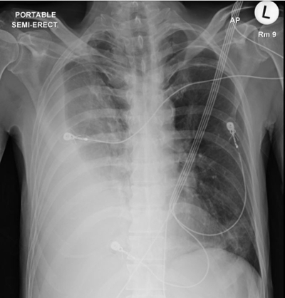
A 56-year-old male with a history of alcohol abuse, was seen in the emergency department with gradually worsening dyspnea. He appeared to be in moderate respiratory distress and his vital signs were normal apart from an increased respiratory rate of 28 breaths/minute and a heart rate of 130. A chest radiograph was performed (image 1)
Images/Video:
Image 1:
Video 1:
Question:
What diagnosis is evident from the ultrasound view?
- Pneumonia
- Simple pleural effusion
- Pulmonary edema
- Complicated pleural effusion
- Atelectasis
D. Complicated pleural effusion
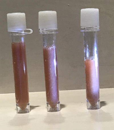
The ultrasound video shows a hypoechoic area in the near-field with extensive septations and dense debris seen in the far field (image 3). This should raise suspicion that the patient has a complicated pleural effusion and potentially needs drainage of the pleural space.
Image 3:
Ultrasound is an extremely valuable tool in the evaluation of the pleural space and serves as an adjunct to physical examination. It helps differentiate non-specific opacities in the lower lung fields such as pleural effusions, pneumonia and atelectasis. It also allows a clinician to localize an anatomical site for thoracentesis with the greatest depth of fluid, thereby increasing procedure success rates and reducing complications such as pneumothorax. (1, 2) Ultrasound can also estimate pleural fluid volume allowing clinicians to track the progression of an effusion without serial chest radiographs. (2)
The appearance of pleural fluid on ultrasound is very important and differentiates simple from complicated pleural effusions. (3) In the absence of debris or septations, a patient likely has a simple transudative pleural effusion. Complicated pleural effusions have an echogenic appearance on ultrasound with or without septations and are more likely to be an exudate. The echogenicity of fluid can also help narrow a differential diagnosis for complicated pleural effusions. Homogenously echogenic pleural effusions are most frequently due to hemothorax or empyema. Effusions with heterogeneous echogenicity suggest a high cellular content related to malignancy. (4) The visualization of septations in an empyema predicts the need for intrapleural fibrinolytics, a prolonged duration of drainage, and the need for possible surgical intervention. (5)
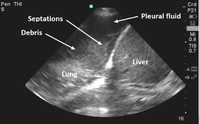
The patient underwent a thoracentesis revealing purulent pleural fluid (shown in image 2). The pleural fluid had an absolute neutrophil count of > 58,000 cells/HPF and was exudative (pleural fluid protein 2.9 g/dL, pleural fluid LDH 7706 units/L, protein ratio 0.73, LDH ratio 33.6) with a pH of 7.00 and an undetectably low glucose. The patient was diagnosed with an empyema and underwent chest tube thoracostomy and subsequent intrapleural fibrinolytic therapy. Methicillin-resistant Staphylococus Aureus grew on pleural fluid culture and the patient was treated with a prolonged course of intravenous antibiotic therapy.
Image 2:
References:
-
Mercaldi CJ, Lanes SF. Ultrasound guidance decreases complications and improves the cost of care among patients undergoing thoracentesis and paracentesis. Chest 2013; 143: 532-538.
-
Balik M, Plasil P, Waldauf P, Pazout J, Fric M, Otahal M, Pachl J. Ultrasound estimation of volume of pleural fluid in mechanically ventilated patients. Intensive care medicine 2006; 32: 318.
-
Yang PC, Luh KT, Chang DB, Wu HD, Yu CJ, Kuo SH. Value of sonography in determining the nature of pleural effusion: analysis of 320 cases. AJR American journal of roentgenology 1992; 159: 29-33.
-
Chian CF, Su WL, Soh LH, Yan HC, Perng WC, Wu CP. Echogenic swirling pattern as a predictor of malignant pleural effusions in patients with malignancies. Chest 2004; 126: 129-134.
-
Chen KY, Liaw YS, Wang HC, Luh KT, Yang PC. Sonographic septation: a useful prognostic indicator of acute thoracic empyema. Journal of ultrasound in medicine : official journal of the American Institute of Ultrasound in Medicine 2000; 19: 837-843.


