Computed tomography (CT) of the thorax (see Figures 2 and 3) showed a markedly dilated esophagus occupying the right hemithorax filled with a large amount of debris, narrowing of the right mainstem bronchus, total atelectasis of the right lung, a large right pleural effusion and leftward mediastinal shift. The patient's gastrointestinal symptoms, along with the dilated esophagus manifested as the air-fluid level on the chest radiograph, are most consistent with achalasia.
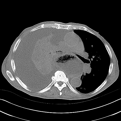
Figure 2. CT of the thorax showing a markedly dilated esophagus occupying the right hemithorax filled with a large amount of debris, narrowing of the right mainstem bronchus, total atelectasis of the right lung and a large right pleural effusion.
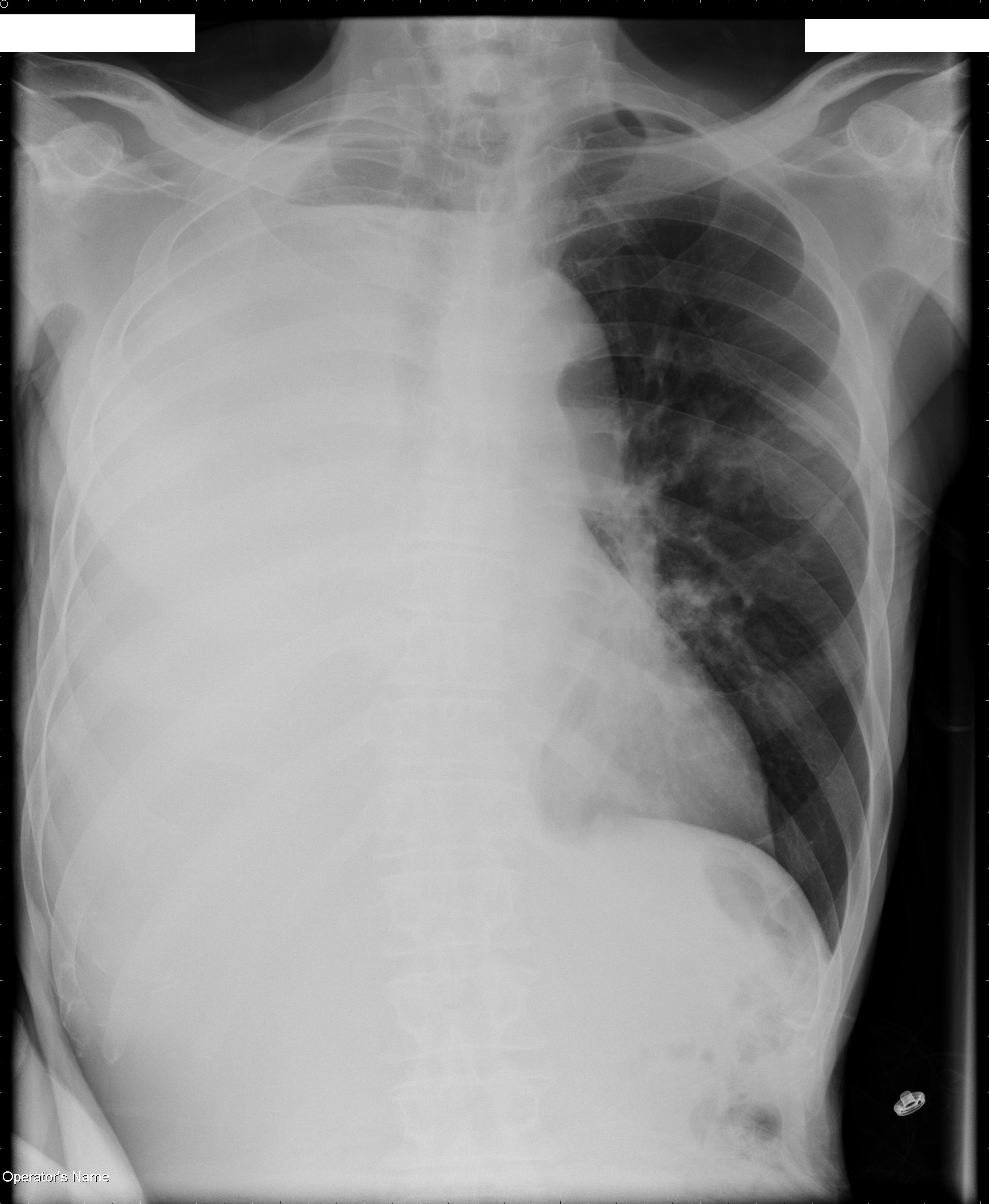
Figure 3. CT image showing narrowing of the right mainstem bronchus (outlined with a white circle) and leftward shift of the mediastinum. Note that an air-fluid level is present in the right mainstem bronchus.
Achalasia is a rare disorder of the esophagus that affects approximately one per 100,000 people. It is characterized by dysfunction of the lower esophageal sphincter (LES) that does not relax appropriately in response to swallowing to allow food to enter the stomach. Patients with achalasia lack inhibitory ganglionic cells in the LES, thereby causing unopposed excitatory signals for sphincter contraction and incomplete relaxation (1). Peristalsis of the lower two-thirds of the esophagus is also dysfunctional, preventing adequate propulsion of food towards the stomach.
Achalasia alone does not explain the radiographic lung findings. Patients with achalasia are at risk for aspiration and, consequently, pneumonia (2). This can result in bronchial obstruction from mucus or aspirated food particles and subsequent atelectasis. Given the patient's history of productive cough and the radiographic findings, his clinical picture is most consistent with achalasia complicated by aspiration pneumonia, total lung atelectasis and parapneumonic effusion.
Hydropneumothorax can cause an air-fluid level on chest radiograph, but in this case, the air-fluid level does not extend to the lateral chest wall, making this diagnosis less likely. Lung tumors with central cavitation can develop an air-fluid level, and lung cancer can certainly cause both bronchial obstruction and malignant pleural effusion; however, a cavitating tumor with an air-fluid level at the right apex would be an unlikely cause of central airway obstruction and total collapse of the right lung.
The major distinction in the diagnosis of a patient with suspected achalasia is between idiopathic achalasia and pseudoachalasia. Pseudoachalasia is most frequently caused by tumor, usually gastric carcinoma, at the gastroesophageal junction. It should be suspected in older patients with a shorter duration of symptoms and significant weight loss. In one series, pseudoachalasia was diagnosed in 9% of patients above the age of 60 with suspected achalasia (3). In idiopathic achalasia, an endoscope should easily pass through the LES; in pseudoachalasia due to tumor, the endoscope should not pass easily, and this should prompt careful inspection and biopsy of the gastroesophageal junction and cardia of the stomach (4).
CT can suggest the presence or absence of a tumor in the distal esophagus but cannot exclude the diagnosis of pseudoachalasia or provide visual or pathologic evidence of malignancy. Likewise, barium swallow can suggest the diagnosis of achalasia by showing a dilated esophagus tapering to the lower esophageal sphincter (the classic "bird's beak" appearance) with loss of peristalsis, but it cannot distinguish between idiopathic achalasia and pseudoachalasia. Esophageal manometry is an important test for establishing the diagnosis of achalasia, characteristically showing aperistalsis in the body of the esophagus and abnormal lower-esophageal sphincter relaxation. However, manometry cannot reliably differentiate pseudoachalasia from idiopathic achalasia. Finally, Chagas' disease, caused by the parasite T. cruzi, is a frequent cause of megaesophagus in endemic regions of Central and South America. Chagas' disease is diagnosed by a compatible clinical picture and confirmed with serologic testing for antibodies to T. cruzi.
Our patient was at risk for pseudoachalasia because of his advanced age, short duration of symptoms and weight loss. EGD revealed a tortuous esophagus, a benign-appearing gastroesophageal junction that was easily traversed by the endoscope, esophagitis and esophageal ulcers. There was no evidence for malignancy at the LES or in the stomach. Esophageal manometry showed aperistalsis of the distal esophagus. Chagas' disease was considered in the differential diagnosis, but because our patient lacked a compatible travel history, serologic testing for Chagas' disease was considered low yield and was not performed. Given the endoscopic findings, lack of evidence for malignancy and supportive results from manometry, the final diagnosis was idiopathic achalasia.
In the presence of total lung atelectasis, lower airway obstruction needs to be excluded. Furthermore, in our patient, aspiration pneumonia was high on the differential diagnosis. Both can be evaluated during bronchoscopy by performing an airway inspection and bronchoalveolar lavage (BAL). In our patient, bronchoscopy showed copious, thick mucus occluding the right mainstem bronchus and external compression of all visualized segmental bronchi on the right. Gram stain of the BAL fluid showed many white blood cells and Gram-positive cocci, while culture grew Mycobacterium fortuitum. This is consistent with previous reports which have suggested that achalasia, complicated by lipoid pneumonia, is a risk factor for nontuberculous mycobacterial pulmonary infections, particularly with M. fortuitum (5). It also highlights the importance of obtaining lower respiratory tract samples for culture to determine the cause of pneumonia in this patient population.
Despite adequate pulmonary toilet and endoscopic evacuation of the esophagus to relieve airway compression, a repeat chest radiograph failed to show re-expansion of the right lung. A thoracentesis was subsequently performed yielding 1600 ml of serosanguinous fluid, leading to partial re-expansion. Analysis of the pleural fluid showed a total protein of 3700 mg/dl, LDH of 97 U/L, glucose of 82 mg/dl, amylase of 106 U/L, white blood cell count of 360/mm3, red blood cell count of 3400/ mm3 and pH of 7.43; cultures and cytology were negative. A chest tube was subsequently placed leading to additional drainage of pleural fluid and further re-expansion of the right lung.
Pleural space evacuation in the setting of airway obstruction could lead to trapped lung, highlighting the need for airway inspection prior to tube thoracostomy. VATS may play a role in the setting of complicated pleural disease, but is more invasive and should be considered if thoracentesis and tube thoracostomy result in treatment failure. Antibiotics alone may have treated infection, but as clinical suspicion for postobstructive pneumonia from aspirated debris was high, airway inspection was indicated to evaluate for this possibility.
Nitrates and calcium channel blockers relax smooth muscle in the LES and thereby lower LES tone. Medical therapy is the least invasive option for patients with achalasia but is frequently ineffective, inconvenient and associated with side effects such as headache and hypotension. Therefore, medical therapy should be reserved for those that are unwilling to perform endoscopic or surgical treatment. Injection of botulinum toxin into the LES inhibits the excitatory neurons that increase LES tone. Short-term relief in symptoms of dysphagia have been seen in up to 50% of patients (6). Pneumatic or balloon dilatation via endoscopy is an effective nonsurgical treatment which can provide at least short-term symptom relief in about two-thirds of patients after a single session (7, 8). Up to one-half of patients, however, will require further therapy within five years with less likelihood of significant symptom relief (9). Surgical myotomy, where the LES is weakened by cutting muscle fibers, can provide similar relief in symptoms compared with pneumatic dilatation with less risk of esophageal perforation (10). Disadvantages to this surgical treatment compared with endoscopic pneumatic dilatation include longer recovery time and frequent development of gastroesophageal reflux postoperatively. A gastrostomy tube, while providing an enteral method of nutrition, will not decrease LES tone or increase esophageal peristalsis.
Endoscopic treatment was recommended to our patient. However, he wished to be closer to his family and was transferred to a university hospital in South Carolina for further evaluation and management. He was lost to follow-up.


