A patient with chest pain and shock
Krittika Teerapuncharoen, MD1, Christopher P. Roth, MD2, Surya P. Bhatt, MD1
1Division of Pulmonary, Allergy and Critical Care
2Division of Cardiovascular Disease
University of Alabama at Birmingham
History of Present Illness: A 73-year-old female presents to the emergency room with a 3-day-history of chest pain and hallucinations. She is initially treated for urinary tract sepsis, however eight hours after presentation, she suddenly became unresponsive and hypotensive, requiring intubation and initiation of vasopressors. Initial focused-bedside ultrasound aiming to evaluate etiologies of shock reveals septal wall hypokinesia.
Past Medical History: Ischemic cardiomyopathy with ejection fraction of 25-30% and placement of a biventricular implantable cardiac defibrillator (ICD), colorectal cancer with recent resection complicated by prolonged hospital stay due to bacteremia and clostridium difficile sepsis.
Physical Exam: She was hemodynamically stable on presentation, without focal neurological deficit. But she developed profound neurologic deficits after episode of hypotension.
EKG: Ventricular paced and no changes from prior EKG. ICD interrogation: No acute events.
Initial labs: Serum troponin of 0.034 ng/mL, serum lactate 3.7 mMol/L, WBC 13.7 x10³ /µL, Pyuria 11-25 WBCs/high power field. Alveolar-arterial oxygen gradient 265 mmHg after intubation.
Imaging: Chest radiograph showed right lower lobe atelectasis with otherwise clear lung fields.
With worsening hypotension, serial bedside ultrasound was performed.
Question: What is the most likely diagnosis?
Impending paradoxical emboli (IPDE)
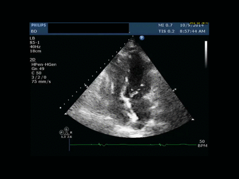
2D transthoracic echocardiogram shows an apical four-chamber view (image A). Visualized is a highly mobile, 2.4 cm free-floating left atrial thrombus and a smaller right atrial thrombus in transit through a patent foramen ovale (PFO). Also noted in the right ventricle is an acoustic shadow that likely represents the ICD wire.
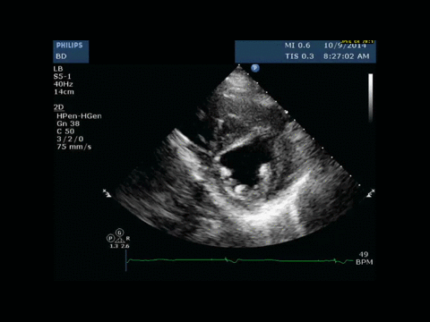
A parasternal short-axis view (image B) shows evidence of right ventricular (RV) pressure overload with systolic flattening of the interventricular septum with severe enlargement of the RA and RV, and severe reduction in the RV systolic function likely from acute pulmonary embolism.1


