A patient with acute onset of chest pain
Michael J. Lanspa, MD
Director of Critical Care Echocardiography Training,
Intermountain Medical Center
Visiting Instructor, University of Utah School of Medicine
History of Present Illness: A 74-year old man presented to the Emergency Room with increasing fatigue over the past week. The day of admission, he had an episode of left-sided chest pain while doing yard work with associated lightheadedness.
Past Medical History: Hypertension, pleural asbestosis.
Physical Exam:
His vital signs were remarkable only for HR 106.
Physical examination was notable only for an elevated jugulovenous pulsation.
EKG: sinus tachycardia with Q wave in lead III.
Initial labs: WBC of 12.3, troponin I 0.26, rest were within normal limits.
Chest x-ray: Notable only for mildly enlarged cardiac silhouette.
An echocardiogram is performed.
Question: What is the most likely diagnosis?
Acute pulmonary embolism.
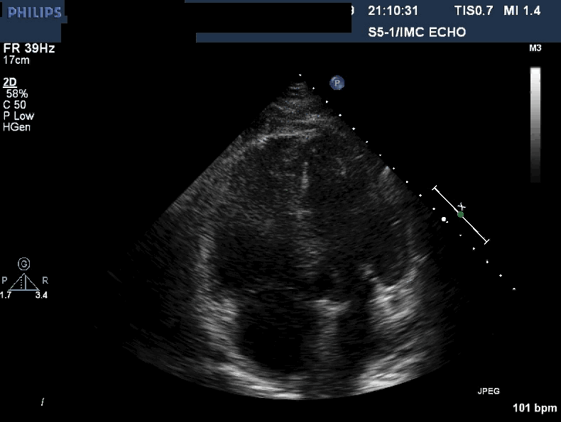
The echocardiogram shows an apical, four-chamber view. This patient has a hyperdynamic, underfilled left ventricle (the chamber on the upper right), and an enlarged right ventricle (chamber on the upper left). The right ventricle is globally hypokinetic, with the exception of the apex, which is hyperkinetic . This hyperkinetic apex is sometimes referred to as an apical wink. This echocardiographic finding (hypokinesia/akinesia in the mid-free right ventricular wall with preserved apical kinesis) is commonly called McConnell’s sign.1
The right ventricle dilates and assumes a more spherical shape, either as a direct response to pressure overload, or due to decreased perfusion of the myocardium from the increased pressure. The mechanism of preserved right ventricular apical function is unclear, possibly related to being tethered to a hyperdynamic left ventricle and/or due to relatively preserved perfusion to the RV apex. This finding was initially thought to be highly specific for pulmonary embolism, but may be present in other causes of acute right heart strain. Most notably, McConnell’s sign may also be present in acute right ventricular infarction.2 Echocardiographic methods that may help differentiate acute pulmonary embolism from RV infarction include estimation of the RV pressures from tricuspid regurgitant jet (lower pressures in RV infarction) and time to achieve peak blood velocity in the pulmonary artery (longer time to achieve peak velocity in RV infarction). In this patient’s case, his RVSP was estimated to be 48mmHg above the right atrial pressure.
Recognition of McConnell’s sign may be particularly useful in a patient with suspected massive pulmonary embolism who is too unstable to go to CT scan, and presumptive diagnosis of pulmonary embolism is needed to justify thrombolytic therapy.
This patient had a saddle embolism seen on CT of chest, and was successfully treated with heparin.
References:


