Orthopnea in a patient with doxorubicin exposure
Jeffrey Halleck, MD
Internal Medicine Resident
University of Utah
History of Present Illness: A 34 year old male with history of Stage 1BX mediastinal B-cell lymphoma treated with 6 cycles of R-CHOP followed by radiotherapy 8 years ago presents with a 2 month history of increasing abdominal girth, cough, dyspnea on exertion, and orthopnea.
Past Medical History: B-Cell lymphoma, chronic pancreatitis, alcoholism, atrial septal defect
Vital Signs: Respiratory rate: 27/min, pulse 112/min, BP 95/60 mmHg, pulse oximetry 91% on 4 liters oxygen via nasal cannula.
Physical Examination: JVP 15cm, bibasilar end-inspiratory rales extending to lower scapular border bilaterally, accessory muscle use, liver span 4cm below costal margin, 1+ lower extremity edema to knees.
EKG: Sinus tachycardia, left axis deviation, left ventricular hypertrophy with repolarization abnormalities.
Chest Radiograph: Findings include a single lead defibrillator, cardiomegaly, prominent interstitial markings bilaterally, central peri-hilar opacities, and mild thickening of the minor fissure.
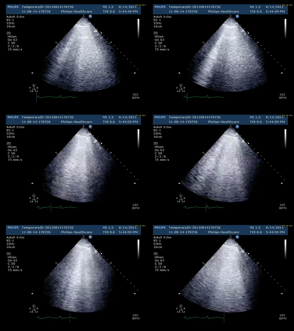
Bedside Lung Ultrasonography: Using either a low-frequency phased-array probe or a high-frequency linear probe, the following finding is present bilaterally on atleast two locationas on each side.

Question: Based on his history and lung ultrasound findings, which one of the following is likely to explain his dyspnea:
- Pneumothorax
- Pneumonia
- Pulmonary edema
- ARDS
Pulmonary Edema
The patient has a new diagnosis of non-ischemic dilated cardiomyopathy induced by prior treatment of doxorubicin for B-cell lymphoma. On 2-D mode ultrasound, there is intact pleural sliding and diffuse B-lines. The B-line is an artifact seen on lung sonography and has the following characteristics:
- It arises from the pleural line
- It is well-defined, and looks like a laser
- It is hyperechoic
- It does not fade
- It erases another artifact called the A lines
- It moves with lung sliding.
The development of B-lines is brought on by accumulation of fluid within the pulmonary interstitium or alveoli causing thickening of the interlobular septum. Interstitial edema first appears within the subpleural interlobular septae. B-lines form as ultrasound beams reflect back and forth as they become trapped in a system involving the pleural line, air-filled alveoli and fluid within the subpleural interlobular septae.2,3,4 The major acoustic impedance of an air-fluid junction creates the long vertical hyperechoic artifact. These artifacts are typically best seen with a low-frequency 3-MHz to 5-MHz phased-array probe, but can also be detected with a high-frequency linear probe.4
The presence of B-lines bilaterally suggests a process known as acute alveolar interstitial syndrome (AIS) which can be caused by ARDS, interstitial pneumonia, pulmonary edema, or any other process involving the pulmonary interstitium (HAPE, ILD, etc.).4 Pulmonary edema is the most common cause for AIS when detected on lung ultrasound. A positive ultrasound study for acute AIS involves visualizing 2 or more B-lines on the screen in more than 2 zones of the lung, bilaterally.3,4 Unilateral B-lines may be seen in pneumonia, lung contusion, or a localized form of acute interstitial edema.2,3 A greater number of B-lines on the screen or shorter distance between B-lines suggest a larger severity of AIS. With worsening edema these eventually become confluent to the point that a diffuse reflective finding known as “white lung” develops. The presence of 3 or more B-lines indicates interstitial edema with 97% sensitivity and 95% specificity.2,3 Chest ultrasonography can be an effective and versatile tool to detect pulmonary edema whether it be detecting decompensated heart failure in an ICU setting or diagnosing and monitoring progression of high-altitude pulmonary edema at a remote high-altitude clinic.1
-
Fagenholz PJ, Gutman JA, Murray AF. Chest ultrasonography for the diagnosis and monitoring of high altitude pulmonary edema. Chest. 2007; 131: 1013–1018.
-
Gardelli G, Feletti F, Nanni A, Mughetti M, Piraccini A, Zompatori M. Chest ultrasonography in the ICU. Respiratory Care. 2012; 57 (5): 773-781.
-
Lichtenstein DA, Meziere GA. Relevance of lung ultrasound in the diagnosis of acute respiratory failure: The BLUE protocol. Chest. 2008; 134: 117-125
-
Lobo V, Weingrow D, Perera P, Williams SR, Gharahbaghian L. Thoracic ultrasonography. Crit Care Clin. 2014; 30: 93-117.


